
|
|
|
Plenary speakers
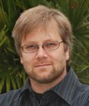 Herwig BAIER, Ph.D., obtained his PhD with Friedrich Bonhoeffer at the Max Planck Institute for Developmental Biology in Tübingen (Germany) in 1995. He then did a postdoc in the laboratory of William Harris at the University of California, San Diego. From 1998 to 2011, he was Professor in the Department of Physiology at the University of California, San Francisco. Since 2011, he is director at the Max Planck Institute of Neurobiology in Martinsried near Munich. The Baier group uses the zebrafish model to understand how neuronal circuits convert sensory inputs into behavioral responses. As zebrafish larvae are optically transparent, transgenic animals that express genetically encoded, fluorescent reporters that indicate changes in neuronal activity can be generated. Coupled with two-photon microscopy, it is possible to watch neurons in the fish brain process visual stimuli and generate motor commands. Additionally, his laboratory pioneered methods for the remote optical control of behavior. This approach involves expression of photoswitchable actuators in select groups of neurons and studying the effects of activity manipulations on CNS network function, sensory perception and behavior. In summary, through his research, he wants to find out how genes assemble neuronal circuits and how these circuits in turn control behavior.
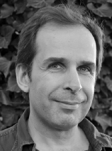 Emmanuel BEAUREPAIRE, Ph.D., is a specialist of optical microscopy of biological tissues. After an undergraduate training in physics in France, he worked two years at Cornell University, and then prepared a PhD in optics at ESPCI ParisTech. He was hired by the CNRS in 2001 and joined the laboratory for optics and biosciences at Ecole Polytechnique (Palaiseau, France), where he initiated the advanced microscopy group. He is now a specialist of multiphoton microscopy. On a methodological side, he develops approaches for removing bottlenecks in live tissue studies, using experimental strategies such as multimodal/multicolor imaging, light-sheet excitation, or wavefront shaping, to address speed/depth/complexity issues. He also explores novel applications fields such as imaging embryonic and nervous tissue development in animal models, and optical diagnostics.
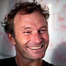 Florian ENGERT, Ph.D., is a german scientist who completed undergraduate work in physics at the Ludwig-Maximilian University of Munich, Germany, and pursued doctoral and post-doctoral studies in neurosciences with Tobias Bonhoeffer at the Max Planck Institute in Munich. He undertook a second postdoctoral post with Mu-Ming Poo at the University of California-San Diego and Berkeley. Florian Engert then came to the Molecular and Cellular Biology (MCB) Department at Harvard University in 2002 and was promoted to Associate Professor in 2006. In 2009, he was promoted to full Professor. While still at the Max Planck Institute, Florian Engert developed a technique for supplementing electrodes with a light-based method for recording from neurons (two-photon laser scanning microscopy). At MCB he has established in vivo electrophysiology and two-photon microscopy imaging in the zebra fish's central nervous system. He also applied the light-activated protein channelrhodopsin-2 to selectively excite neurons and gage their effect on zebra fish behavior. Combined with functional imaging and behavioral assays, these techniques allow him to monitor and control the underlying neuronal activity that induces fish to respond to certain stimuli and then explore how those stimuli are processed in the brain and how they change behavior.
 http://labs.mcb.harvard.edu/engert/
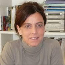 Valentina EMILIANI, Ph.D., obtained her PhD in Physics at the University ‘La Sapienza’ in Rome (Italy) in 1996. She was working on the investigation of tunneling effect in asymmetric double quantum wells by ultrafast spectroscopy. From 1997 to 2000 she joined the group of Prof. Thomas Elsaesser at the Max Born Institute of Berlin as a post doc, working on the investigation of carrier transport in single quantum wire by low temperature scanning near field optical microscopy (SNOM). From 2000 to 2002 she was in the laboratory directed by Prof. Marcello Colocci at the European laboratory for nonlinear spectroscopy, Florence Italy. There she worked on the investigation of light propagation in disordered structure by SNOM. From 2002 to 2004 she was at the Institute Jacques Monod, where she was working on the investigating of the role of mechanical forces on the establishment of cell polarity by the use of the optical tweezers technique. In the year 2005 she obtained the price EURYI 2005 and she moved in the Neurophysiology and New Microscopies Laboratory where she formed the “Wave front engineering microscopy” group. The group has pioneered the use of wave-front engineering for neuroscience by demonstrating a number of new techniques for efficient photoactivation of caged compounds and optogenetics molecules, techniques based on computer generated holography, generalized phase contrast and temporal focussing. In 2014 she has been appointed Director of the Neurophotonics Laboratory at the university Paris Descartes.
http://www.neurophotonics.parisdescartes.cnrs.fr/
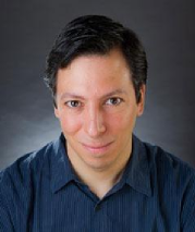 Randy BRUNO, Ph.D., obtained his PhD at the University of Pittsburgh School of Medicine under Daniel Simons’ supervision and completed a postdoctoral fellowship at the Max Planck Institute for Medical Research with Nobel Prize-winning Bert Sakmann. He is currently Associate Professor of Neuroscience at Columbia University. With Sakmann, he developed a way to measure the strength of a connection between a cell from the thalamus – which relays input from a sensory organ (a whisker or an eardrum) – and a cortical cell in a living animal. Using this technique, his own lab recently showed that thalamus accounts for all the sensory-driven activity of deep cortical layers. The focus of Professor Bruno's present research is how cortical circuitry mediates cognition, particularly sensation. Despite their diverse functions, the many distinct areas of neocortex have the same cell types arranged in the same laminar structures, and having the same general connectivity with each other and with other areas of the brain. It is as if nature reiterated this one circuit for many different tasks. His goal is to reverse engineer that circuit. He is pursuing this goal by imaging behaving transgenic mice using two-photon and single-photon microscopy techniques. The Society for Neuroscience presented the Young Investigator Award to Randy Bruno in 2013.
 http://www.neurosciencephd.columbia.edu/profile/rbruno?profile=researcher
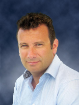 Mickael TANTER, Ph.D., is Research Professor of the French National Institute for Health and Medical Research (INSERM). For eight years, he is heading the Inserm laboratory U979 “Wave Physics for Medicine” at Langevin Institute, ESPCI Paris, France. His main activities are centered around the development of new approaches in Wave Physics for medical imaging and therapy. His current research interests a wide range of topics: Elastography using Supersonic Shear Wave imaging, Ultrafast ultrasound imaging and more recently the concept of fUtrasound (functional ultrasonic imaging of brain activity) he introduced with his team. He is the recipient of 26 world patents in the field of ultrasound and the author of more than 170 peer-reviewed papers and book chapters. In 2006, he co-founded Supersonic Imagine with M. Fink, J. Souquet, C. Cohen-Bacrie. Supersonic Imagine is an innovative French company positioned in the field of medical ultrasound imaging and therapy, that launched in 2009 a revolutionary Ultrafast Ultrasound imaging platform called AixplorerTM with a unique real time shear wave imaging modality for cancer diagnosis (>140 employees, 102 M€ venture capital, and more than 800 ultrasound systems already sold worldwide). He received many scientific prizes and was recently awarded a 2.5 M€ European Research Council (ERC) Advanced Grant to develop the concept of fUltrasound for clinics and neuroscience research ( http://www.fultrasound.eu/ ).
 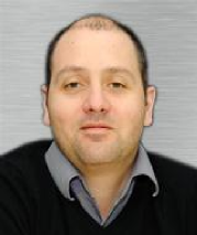 Frederic LESAGE, Ph.D., did his PhD in Physics at the Paris-Sud University after a Master of Science in applied mathematics and physics at Cambridge University. Now a researcher at the Montreal Heart Institute (MHI) and a professor in electrical engineering at the École Polytechnique de Montréal since 2005, he specializes in the development of optical imaging techniques. Before coming to Polytechnique, he studied and held industry positions in bioimaging. He currently manages a laboratory of 20 people. His research activities are dealing with the development of novel imaging techniques for neural imaging. These techniques are used with both, humans (diffuse optical imaging of the brain during cognitive tasks and study of the neuronal metabolism) and small animals (study of neuro-degenerative diseases by means of transgenics and molecular fluorescent probes). He is particularly interested in the question of brain and vascular aging and performed a number of studies on brain aging using various optical imaging techniques such as diffuse optical tomography, near-infrared spectroscopy, optical coherence tomography and two-photon microscopy.
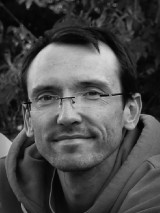 François ROUSSEAU, Ph.D., got his PhD in 2003 at the Rennes 1 University. He then went to San Francisco University (UCSF) for a post-doctoral contract working on MR spectroscopy brain atlas building and image reconstruction of in utero fetal brain MRI. Since 2006, he is a CNRS research scientist in an image processing laboratory currently named iCUBE, at Strasbourg University (France). François Rousseau's works are in the frame of an interdisciplinarity program that aims at providing biomedical image processing tools for diagnosis and longitudinal studies. He is currently working on analyzing and simulating the cerebral development of fetuses and infants using anatomical and diffusion MR imaging. The main aim is to understand the relationship between cerebral changes and cognitive development.
 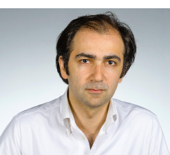 Alipasha VAZIRI, Ph.D., is a physicist and currently a group leader at the Institute for Molecular Pathology (IMP), Associate Professor at the University of Vienna and the director the Interdepartmental Research Platform Quantum phenomena and Nanoscale Biological Systems (QuNaBioS). He obtained his PhD in the field of quantum optics at the University of Vienna with Anton Zeilinger in 2003 and continued as a post-doc with Dr. William Philips at the National Institute of Standards and Technology (NIST) in Maryland, USA. In 2007 he moved as a research specialist to Howard Hughes Medical Institute, Janelia Farm Research Campus where he focused on the development of new optical techniques, such as super resolution microscopy and patterned optogenectis to study biological structure and function at high speed and resolution. In 2011 he moved as a group leader at Institute for Molecular Pathalogy (IMP) and Assistant Professor at the University of Vienna to Austria. His research program is targeted at developing and using optical tools to understand how the dynamic of large neuronal networks is related to sensory inputs and behavior. Amongst others his lab has pioneered the development of optical techniques that allow high speed single cell specific application of arbitrary optogenetic patterns on large neuronal populations and high speed parallel readout of neuronal activity at the whole-brain level.
 | Online user: 1 | RSS Feed |

|
